The use of two different nanoscale or atomic scale measurements to analyze the same sample is critical to mastering the properties of a material or product. This complementary analytical approach is particularly useful for measuring new generations of nanoscales such as MOSFETs and FINFETs. The doping profile of the transistor. In this paper, two methods of electron tomography (ET) and atomic probe tomography (APT) were used to study the spatial distribution of boron atoms in nanoscale transistors. In this paper, the electron tomography technique is combined with atomic-scale resolution ion bombardment simulation to correct the three-dimensional reconstructed image distortion of atomic probe tomography. APT 3D reconstruction technology enables detailed chemical analysis of sample devices.
In nanoscale transistors on very large scale integrated circuits, especially in complementary metal oxide semiconductor (CMOS) devices with increasingly reduced external dimensions, the atomic spatial distribution of dopant species is a major problem. The gate voltage value at the time of the strong inversion layer transition on the MOSFET channel is called the threshold voltage. The threshold voltage change is an important characteristic related to the atomic number, spatial distribution and properties of the dopant. 3,4, 5 . Therefore, understanding the doping spatial distribution is of great significance for the development of advanced technology nodes, and it is necessary to accurately determine the spatial distribution of doping on the submicron scale. In addition, to analyze small-sized CMOS transistors, it is necessary to use a characterization method that can perform three-dimensional reconstruction and quantitative analysis of the doping profile.
Atomic Probe Chromatography (APT) 6 is currently recognized as the primary measurement and analysis method for atomic spatial 3D reconstruction of selected small (100x100x1000 nm3) chemical samples at the atomic scale. APT is one of the main tools for microelectronics companies to analyze current and future semiconductor components. As a quantitative microanalysis method, it can find light and other light elements such as boron in silicon. The rapid development of laser APT technology plays a decisive role in the study of 2D 11 and 3D 12, 13 structures in the 9th and 10th circles of nanoscience. Recently, in the process of analyzing the doping profile of the gate and sidewall of a FinFET structure using the APT method, we have found undesired characteristics in the doping process 14,15. In addition, the APT experimental results show that the channel region doping concentration profile of each MOSFET device under test is related to the observed threshold voltage distribution of the MOSFET device.
APT is a unique method for microelectronic measurement analysis, but this method is subject to many limitations when measuring three-dimensional structures composed of different evaporation field regions (eg, different chemical compositions and/or crystal structures). The low (high) change in the evaporation field (F) causes the radius of curvature (R ~ V/F) to become smaller (larger), resulting in local focus (defocus), which causes the detector to be affected by high (low) density. Therefore, from the three-dimensional reconstructed image, the low evaporation field (for example, the metal gate and channel of the transistor) appears to be a high atomic density region. This local amplification phenomenon not only causes image distortion 17-21, but also causes ion trajectory. An overlap occurs near the phase boundary, causing the component data to shift. To fix images and eliminate artifacts, the industry has developed simulation and complex reconstruction methods. The cause of image distortion is that the ion trajectory is offset near the phase boundary 22, 23. Density repair methods work well (density relaxation along the xy and z axes) when image distortion or distortion is not apparent. However, when the evaporation fields between the phases are different, the repair method is difficult to work. These problems become prominent when analyzing complex structures such as MOSFET transistors. Complementary microscopy with integrated electron tomography (ET) and atomic probe chromatography (APT) methods minimizes these problems when analyzing nanoscale MOSFET transistors. Electron tomography makes it possible to characterize device structures at nanoscale resolution using 3D images 24, 25 without pinpoint damage and body image distortion. The advantage of this dual analysis method is that APT and ET analysis methods can be used to analyze the same object. This paper describes how to use the electron tomography (ET) and atomic probe tomography (APT) probe tomographic methods to quantify the lowest distortion rate of the boron atomic spatial distribution of a 45 nm gate length PMOS transistor. These experimental results demonstrate that complementary microscopy is beneficial for engineers to understand and improve advanced nanoscale semiconductor devices.
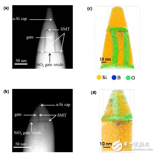
This paper explores two dedicated MOST transistors for the 45 nm gate test structure, using stress memory technology (SMT) to introduce stress into the channel. A polysilicon film is deposited on the 3 nm thick SiO2 layer, and after etching, a polysilicon gate structure is formed, and then boron atoms are implanted, the incident energy is 2 keV, and the dose is 1.1×10 15 at.cm-2. At this time, the periphery of the gate is a SiO2 SMT layer. The device is annealed to diffuse and activate impurities in the gate region and source/drain regions. Finally, an amorphous cover (a-Si) is deposited over the trench and gate structures. The difference between the two transistors is the difference in geometry: the first device, DEVICE1 [Fig. 1 (a)], has a gate length of 45 nm and a gate height of 100 nm, while the second device, DEVICE 2 [Fig. 2(b)], has a gate length and gate. The heights are 90 nm and 50 nm, respectively. Because of the different sizes, the geometry of DEVICE1 is similar to that of a FINFET transistor. It should be noted that the gate of DEVICE2 is too long and only a part of the gate is included in the tip, as shown in Figure 1.b. The ET and APT dual analysis requires the use of needle-shaped samples, which avoids shadowing effects during tilt acquisition and promotes field evaporation at the tip of the tip. The tip is prepared by first thinning the sample with a FEI Strata milling device and taking the sample by in-situ lift-out, followed by circular milling of the Ga ion with 30 keV, followed by 2 keV. Low energy cleaning process.
The electron tomography experiment was performed in a HAADF-STEM mode using a FEI 80-300 kV Titan microscope with an operating voltage set at 200 kV. In order to keep the sample tilted, a dedicated APT probe-compatible tomography mount (Fischione 2050 coaxial rotary tomography mount) was used. The probe has a convergence angle of 5 mrad, a probe size of 0.33 nm, and a field depth greater than 80 nm. To obtain the tilt sequence, the tilt angle range is set from -78° to +78° and the tilt step is 1°. The projection alignment uses a cross-correlation alignment algorithm, and the volume reconstruction uses the FEI Inspect 3D software with built-in synchronous iterative reconstruction algorithm. Atomic probe tomography was performed on a FlexTAP instrument (CAMECA) using a magnified erbium doped element laser with a wavelength set at 343 nm, a duration of 450 fs, an emission energy of 35 nJ/pulse, and a repetition rate of 100 kHz. During the analysis, the tip temperature was maintained at approximately 70 K.

Figure 1. HAADF STEM image of DEVICE1 (a) HAADF STEM image of DEVICE2 (b) APT and ET analysis requires the sample to be prepared into a needle shape. APT body diagram of DEVICE1 (c) APT body diagram of DEVICE2 (d) A dot represents an atom. Note: Because the gate oxide/substrate interface has a tip break, DEVICE1 is always analyzed to the gate oxide.
Figure 1(a) shows the HAADF STEM image of the low magnification DEVICE1 with the region of interest intact and in the middle of the tip. The dark contrast and the bright contrast are assigned to the SiO2 and silicon regions, respectively. SiO2 under the polysilicon gate is a gate oxide region, and SiO2 at the periphery of the gate corresponds to an oxide layer. The source and drain regions are also indicated in the figure. Figure 1(b) shows a HAADF STEM image of a partial structure of DEVICE2, including a source/drain region and a portion of the gate and silicon oxide. Figure 1(c) shows the APT three-dimensional atomic distribution of the gate region of DEVICE1. Unfortunately, the tip of the tip fails in the gate oxide layer and the source/gate region image cannot be acquired. This volume reconstruction uses the standard tip distribution method 26, 27. As expected, boron atoms were found in the polysilicon gate region, but boron atoms were also found in the SMT SiO2 layer. To quantify the spatial distribution of DEVICE2 source/drain boron doping, another configuration consisting of a partial structure of DEVICE2 was used. Figure 1(d) shows the three-dimensional spatial distribution of oxygen and boron atoms in the gate and source/drain regions of DEVICE2, consistent with the doping implantation process. In addition, compared with HAADF images, APT reconstructed images have distortion and size shift problems. For example, both devices under test have oxide layer amplification and silicon gate region reduction. These image deformation phenomena can affect the quantitative analysis of boron atoms.
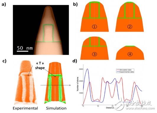
The local amplification phenomenon is particularly noticeable on the DEVICE1 with a short gate length. The silicon gate appears to be a compressed layer (two-thirds to three-quarters smaller), while the oxide layer is significantly amplified (three to four times magnification). Complex system experiments have shown that ion ballistic overlap is not only related to field evaporation, but also to the shape and aspect ratio of the object of interest. For example, it is currently known that the composition data offset is very significant near the small precipitation interface, and therefore, a component data correction process 28 is required. These effects 29, 30 can be observed in systems consisting of silicon embedded in a SiO2 matrix, and the interfacial mixing thickness is estimated to be approximately 2 nm. It has been found that there is also interfacial mixing between the Si and SiO2 phases within the Si/SiO2 layer structure. In addition, when the field evaporation between small deposits is extremely different and the matrix is ​​present, ion defocusing in the particles is expected to cause component mixing. For the three-dimensional complex architecture of DEVICE 1, it is not enough to briefly describe the local amplification effect. In fact, field evaporation and lens reconstruction caused by complex geometric tips are an unsteady phenomenon. The primary oxidation zone of the gate region first appears as a large cubic SiO2 phase embedded in the silicon on the surface of the tip; then, it gradually transitions into a Si/SiO2/Si/SiO2/Si film structure parallel to the tip of the needle, and finally forms a A SiO2 gate oxide layer perpendicular to the tip of the needle. Therefore, we should thoroughly understand the impact of this complex structure on reconstruction. To understand the root cause of APT reconstructed image distortion, the process of atomic evaporation of surface atoms containing DEVICE1 must be simulated. Although the device size allows for actual 3D simulation, it is cumbersome to operate. Taking into account the evaporation fields of the silicon and SiO2 layers, a cylindrical symmetry method is used to simplify the simulation model, and a detailed description of the simulation process can be found elsewhere. These semi-quantitative simulations provide evolutionary characteristics of tip shape over time during field evaporation and calculate ion trajectory and impact locations, which are then used to create simulated three-dimensional images using standard image reconstruction algorithms. Obviously, limited to the cylindrical symmetry assumption, the simulation model cannot be expected to quantify the local amplification effect. However, continuous changes in geometry and phase composition during field evaporation must not be violated, and the main evaporation characteristics and experimental results images can be compared [Fig. 2a]. In addition, the simulation model can also simulate the actual size structure within a reasonable simulation time. The value of the silicon evaporation field (33 V/nm) is known, and the value of the SiO2 evaporation field is unknown. When crossing the Si/SiO2 interface, the evaporation field can be calculated from the threshold pressure using the Jeske and Schmitz32 methods. Our system measurements give an estimate of the field evaporation of SiO2 (43 V/nm), which is about 25% higher than the silicon substrate.

Figure 2. A) Simulation configuration of the device. The STEM image size is scaled up. b) Tip shape change for different evaporation stages c) Reconstructed image of the oxidation zone around the silicon gate as an experimental and simulated object. The defocusing effect caused by the high evaporation field of the SiO2 phase causes the thickness of the oxidation zone to become large. Note: The T shape above the gate region below the SMT oxidation region is the result of these partial amplifications. Because the image is distorted, no magnified image is provided. d) The horizontal distribution of the density of the silicon gate center. The low density zone is associated with the oxidation zone (green in Figure 2c).
Figure 2(b) shows the simulation results of DEVICE1 in different field evaporation stages. There is a high evaporation field on the SiO2 phase, which causes a significant difference in the local radius of the upper part of the silicon gate region. In the first stage of field evaporation, the closed SMT SiO2 layer around the silicon gate region appears to protrude from the tip surface, resulting in enhanced amplification, which causes the SiO2 SMT layer region to appear amplified on the detector. When evaporating under the silicon gate phase, the surface of the tip has a tendency to be flat or close to a negative curvature, and as the amplification effect rapidly decreases, the upper portion of the gate forms a "T" shape (Fig. 2.c). This rapid change in the tip axis is concentrated only on the ion trajectory and causes projection distortion [Fig. 2(c)]. Figure 2(d) shows the atomic density distribution from the device field evaporation simulation. Except for the low density seen in the center of the volume map, other simulation results are consistent with the image deformation analysis in the experiment. Therefore, the atomic density ratio of the silicon phase to the SiO2 phase in the device experiments and simulations was 10. The reason for the low-density area on the tip axis is that the simulated model system has a low-index crystal. Because of the polysilicon properties of the gate, no low density regions were present in the device experiments.

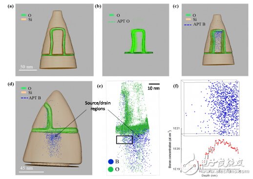
Figure 3: (a) Three-dimensional reconstructed image of DEVICE1 after segmentation (b) Mercury reconstructed volume map of atomic probe chromatography (dashed line) and oxygen reconstructed volume map of solid tomography (solid line) (c) Three-dimensional APT map of boron distribution in the gate region (blue dotted line) (d) Three-dimensional map of boron atom distribution in the gate region, channel and source/drain regions of DEVICE2 (e) Density correction APT pattern (f) groove of DEVICE2 The 1-D boron concentration profile in the channel (corresponding to the selected atomic distribution illustration).
Except for significant image distortion in both simulation and experimentation, the simulation model did not foresee large-scale atomic mixing. Therefore, data reconstruction can be improved by density relaxation methods. After using a method 33 based on atomic density correction, the artifact phenomenon is reduced in the first order, and this method does not affect the local components. By matching the electronic tomographic volume data with the modified APT volume data, we validated the improvement of APT reconstruction using an iterative algorithm. This method can find the best parameters for APT reconstruction. Fig. 3(a) shows a three-dimensional reconstructed image of the surface rendering of DEVICE1 derived from an electron tomography study. The body strength segmentation method uses the absolute threshold method. Therefore, the oxidation zone body map and the silicon region body map are not difficult to distinguish, and are represented by green and orange, respectively. Therefore, it is feasible to provide a complete three-dimensional characterization of the device. Figure 3(b) shows the oxygen reconstruction of the electron tomography oxygen-reconstructed body (green solid line) and density-corrected atomic probe chromatography. A merged image of a volume map (green dotted line). As shown in Figure 3(c), this dual analysis method improves the authenticity of the atomic profile of the three-dimensional reconstruction of boron throughout the device by atomic probe chromatography after characterization using electron tomography. For larger devices other than DEVICE1, we have determined a more complete boron doping profile, as shown in Figure 3(d), where the three-dimensional distribution of boron atoms at source/drain, channel and gate The same is the dual approach of ET and APT. As shown in Fig. 3(e), the distribution of boron atoms in DEVICE2 is not offset, and the 1-D boron concentration of the source/drain and channel regions is calculated, and the maximum boron concentration of 2.7×10 20 at.cm-3 is calculated. The doping conditions are the same. The detection threshold is estimated to be 3 x 10¹⁹ at. cm-3, which is limited by the background noise in the mass spectrum. The source/drain channel boron profile shows a lateral gradient of 5 nm/decade, which indicates that the annealing process results in lateral diffusion of boron atoms, which is consistent with the observed phenomenon in doped FINFET 14.
In summary, simultaneous analysis of the same device using electron tomography and atomic probe chromatography can correctly measure the boron-doped spatial distribution of the gate and source-channel regions of nanoscale devices. The electron tomography image directly shows the image distortion in the APT volume map, which can interfere with the boron quantification analysis. We performed a three-dimensional numerical encoding method with cylindrical symmetry to simulate atomic evaporation of a device similar to the FinFET configuration. The simulation results show that the high evaporation field of the SiO2 SMT layer causes the silicon gate region to be compressed, which is the same as the experimental results here. This shows that it is important to use the electron tomography method to better evaluate, understand, and correct the image distortion caused by the field seen in complex structures. The density correction algorithm, combined with electronic tomography and atomic probe volume data iterative matching, can minimize APT 3D projection distortion. APT is the only analytical method that can quantify elements in complex structures in three dimensions, extending the range of measurement and analysis using electron tomography alone. This powerful dual analysis method helps analyze the doping profile of next-generation devices such as FinFETs to better understand and improve the electrical characteristics of these devices.
Litecoin (LTC) is a cryptocurrency created as a fork of Bitcoin in 2011. It uses a hashing algorithm called Scrypt that requires specifically designed mining software and hardware. It is minable, and continues to rank in the top cryptocurrencies for value and trading volume.
Litecoin mining is the process of validating transactions in the blockchain, closing the block, and opening a new one. Litecoin uses the proof-of-work consensus mechanism, which uses computational power to solve the nonce, which is part of the hash, that secures the block. The hash is the alphanumeric sequence of numbers that is encrypted by the hashing algorithm. When the nonce is solved, Litecoin is rewarded.
Litecoin mining became popular in 2011 when Charlie Lee, a software engineer at Google, announced its creation as a Bitcoin fork with modifications intended to help it scale more effectively.
Just like Bitcoin, it can be mined on computers using central processing units and graphics processing units. However, it isn't as profitable or competitive as purchasing an application-specific integrated circuit (ASIC) and joining a mining pool.
Ltc Asic Miner,Antminer L3 Plus Plus,Bitmain Antminer L3 Plus,Bitmain L3 Plus
Shenzhen YLHM Technology Co., Ltd. , https://www.ylhm-tech.com